


Radiology QA & Dosimetry
Please click the product you want to learn more about:
Enquiry Now
Diagnostic Radiology
i. Beam Measurement
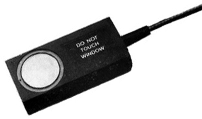
QUART DidoCT
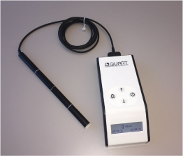
Pencil Chamber Meter for CT Applications
The QUART didoCT meter is designed for easy and precise dose-width product measurements.
The meter does not require any pre-setting procedure for direct reading of DWP, rate and time parameters. Its detector part is based on solid-state technology. Unlike conventional ion chambers, the QUART didoCT is not affected by variations in environmental temperature or air pressure and does not require correction.
The didoCT is equipped with a backlit display to assure swift readings even in darkened environments. To provide the ability to track generator characteristics, the dose or DWP rate is refreshed continuously on the meter display while the measurement is running.
The meter is powered by a rechargeable battery. One charge is sufficient to last approximately 80 hours of continuous use. Recharging the meter until full takes only between 3–4 hours. A warning will appear on the display when the battery charge is running low.
Additional Features
- Measures kVp, kVeff, mA, mAS, exposure time, dose, HVL
- Easy to use
- kV measurement is non-invasive
- AC or DC x-rays
- High accuracy
- mA invasive or non-invasive (option)
- Two year warranty
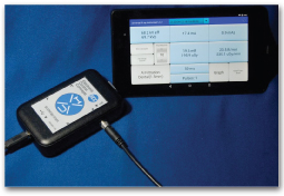
USB Tablet Display
- 7 inch Tablet display for reading outside x-ray room
- Tablet holds stored readings
- Export data to spreadsheet
- Email or transfer data to computer
- Display graph of X-ray waveform
Typical Applications
- Verification of peak X-ray voltage
- Constancy checks
- Calibration of X-ray
- Quality assurance
- Measurement of exposure time
- Half value layer determination
- Troubleshooting and repair of X-rays
- Maintain quality of X-ray
Model UXI
Kvp, mAs, Dose & Exposure Time Meter
- kVp measures the effective and peak x-ray accelerating voltage from tungsten x-ray generators
- mA measures x-ray tube current
- Non-invasive current clamp option
- Dose measures the radiation output over an area
- Works with dental, radiographic and fluoroscopic x-rays
- Place unit in x-ray beam to measure kV, dose, dose rate, Half-Value Layer (HVL), pulses, and exposure time. See readout and graph on tablet.
- Comes with protective carry case
Special Feature: CT-kV Measurement
As an optional feature, the QUART didoCT can be supplied with free-in-air kV measurement capability.
The meter’s kVp feature is designed to non-invasively measure the generator output. It is calibrated at suitable standard radiation qualities in accordance with the majority of computed tomographs used in radiology or radiation therapy.
Model 870
mA, mAs & Exposure Time Meter
The Model 870 is a solid-state, digital instrument designed to assess the performance of medical X-ray generators. This instrument measures and displays mA, mAs, and exposure time for each X-ray exposure and can be used to test dental, radiographic, and fluoroscopic X-ray units. This is a new state of the art instrument with many advanced features. For example, entering correction factors for different types of X-ray waveforms is not required since the Model 870 has the capability of automatically determining the type of X-rays being measured. In addition mA, mAs and exposure time are displayed and stored for each X-ray exposure further minimizing the number of X-ray exposures.
Model 8000 NERO®mAx
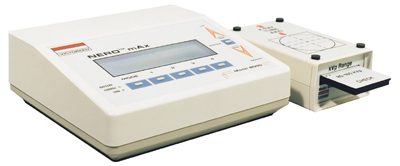
Non-Invasive X-Ray Beam Analyzer
The highly successful NERO® (Non-invasive Evaluator of Radiation Outputs) tradition continues with greater accuracy and more functions than ever. The Model 8000 NERO®mAx is a fifth-generation instrument featuring unprecedented a 100kHz beam sampling rate and direct mA/mAs measurements. The NERO®mAx’s innovative EFM (Easy Flow Menu) system and flexible soft keys provide an intuitive, user-friendly operating environment for quick, accurate, and easy measurements.
The compact and simple control console houses the rechargeable battery, display, 8 control buttons, and the sophisticated electronics for accurate, easy to read, reproducible measurements. All measurement modes are displayed on the super-bright, backlit display and are controlled by the 5 soft keys directly below and 3 hard keys to the right of the display. Connectors for the power input, RS-232, printer, scope output and remote detector are located on the rear panel of the console.
The NERO®mAx detector module contains sensors for simultaneous measurement of kV, exposure or rate, and mA or mAs. An ionization chamber, located immediately below the top surface of the detector module, is used for exposure/rate measurements. A variety of external ion chambers may be connected for specialized in-air or in-phantom exposure measurements. Chamber calibration factors can be stored for direct readout in R or Gy.
KV is measured by a set of solid-state detectors located below the ion chamber. Differential absorption is accomplished by removable filter cards. Each filter card is coded for proper insertion, identification of which filter set is in position and verification of its validity for the selected measurement mode. The W/Al filter card and the Mammo filter card are clearly labeled as to the x-ray targets for which they are calibrated. The Mammo filter card includes the following target/filter combinations: molybdenum/molybdenum, moly/rhodium, moly/aluminum, rhodium/rhodium and rhodium/aluminum.
Time is accurately calculated by NERO®mAx from the number of sample data points collected during the entire exposure. mAs is measured invasively, using two test leads provided with the NERO®mAx. The NERO®mAx Excel Add-in software imports measured data and waveforms directly into an Excel spreadsheet to maximize flexibility for report generation. The NERO®mAx basic system consists of the control console, detector module, 2 filter cards, 2 mAs leads, Excel Add-in, AC adapter, 3 aluminum HVL plates, operator manual and carrying case. CNMC can supply a NERO®mAx system that includes a Pentium® notebook PC, Excel spreadsheet software, a variety of external ion chambers, phantoms and other accessories.
Model 4000M+
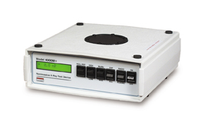
Non-invasive X-Ray Beam Analyzer
The Victoreen 4000M+ measures kVp, exposure and time simultaneously and non-invasively. In addition to its ability to make accurate measurements on tungsten/ aluminum tubes, it is also capable of performing kVp, dose and time measurements on molybdenum anode mammography tubes. An external ion chamber connector provides an interface to a variety of external ionization chambers.
Operation of the 4000M+ is simple and straightforward. The operator simply places the instrument, with the switches set appropriately, on the X-ray table and makes the exposure. The display automatically updates, sequentially displaying the measured values. The instrument resets automatically, being instantly ready to take another exposure. Measurement data can be transferred to a personal computer using Microsoft® Excel Add-In software.
Five user-selectable filter pairs ensure optimum accuracy over the entire diagnostic range, with minimum filtration dependence. Exposure measurements are made with an integral plane-parallel ionization chamber, located above the filter wheel. Exposure time is measured with quartz crystal accuracy.
Model 06-526 RAD-CHECK® PLUS
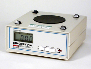
X-Ray Exposure Meter
The Model 06-526 RAD-CHECK® PLUS is a rugged, miniaturized system for making routine checks of X-ray exposure and exposure rate. Though priced much lower than equipment producing comparable results, its reliability, accuracy and ease of operation warrant its use in any QA program. This compact instrument consists of an electrometer with display electronics and a built-in ion chamber in one compact package.
Operation is simple: Just place the instrument on the X-ray table, collimate the beam to the detector using the visible light beam, make an exposure and read the results.
Applications
Fluoroscopy Exposure Measurements
These are needed to check equipment calibration, operation of the automatic brightness system and to determine that the maximum exposure rate is within the legal limits.
Exposure Checks – Radiographic (mR/mAs)
Regular radiography output checks to assure that the radiation output has not changed due to aging, drift, component failure or miscalibration.
Beam Quality – Half Value Layer (HVL)
The beam quality check determines whether the filtration is at least the minimum required by law. This filtration is needed to reduce patient exposure from the low energy portion of the X-ray energy spectrum.
mAs Reciprocity – mA Station Checks
At a given kVp, the mR/mAs values obtained should be reproducible and independent of particular values of mA and time used.
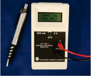
Model 815
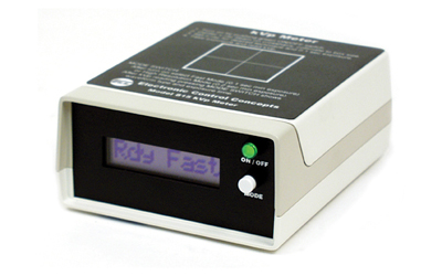
Digital kVp Meter and Exposure Timer
The Model 815 is a versatile, multipurpose digital kVp meter, optimized for dental X-rays but also usable on conventional and fluoroscopic X-rays. In addition, it measures the time duration of X-ray exposure.
Operation can be learned in minutes: simply place the Model 815 in the beam and make an exposure. The large display is easily readable from outside the X-ray room and the alphanumeric messages provide easy to understand operational and diagnostic status.
Analog waveform output for an oscilloscope is provided for waveform analysis – half wave, full wave, DC and 3 phase.
Model 8700
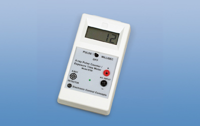
X-Ray Pulse Counter & Exposure Time Meter
The Model 8700 is a versatile digital X-ray pulse counter and exposure time meter. It measures the time or duration of radiation output produced by a wide variety of X-ray generators.
For direct measurement of exposure from the X-ray head, simply place the 8700 under the head and make an exposure. The 8700 is “self-resetting". There is no need to reset the instrument after each reading. The reading is stored after each exposure, until the next exposure.
When testing X-ray timers and controls, the time of the relay contact closure can be measured using the AC input feature. When used with DC, capacitor discharge and 3-phase X-ray, exposure time is measured in milliseconds.
Typical applications include: verification of X-ray exposure time, calibration of X-ray timer, quality assurance, measurement of exposure, direct measurement of timer accuracy and analysis of X-ray malfunctions.
An optional remote sensor is available to allow use of the 8700 at distances of up to 12 feet away from the X-ray machine.
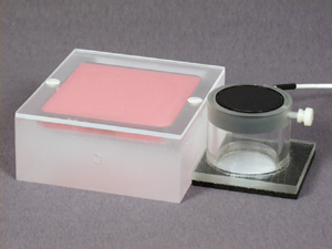
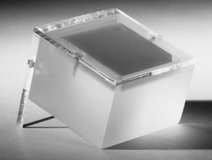
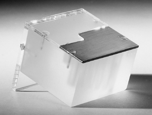
QUART DIDO Series
DIDO2000K & DIDO2100K
Compact Design Concept
The DIDO series diagnostic dosimeters are multifunctional Quality Assurance platforms. Strictly following our own Compact Design Concept, they feature optimised size and design plus a compact multi functional state-of-the-art detector.
Downsize-detector design facilitates measurements where only limited space for a proper detector positioning is available. Hence, measurements behind the scatter radiation grid of a radiography unit can be done with the DIDO without any limitations. And, no influence whatsoever is exerted on the automated exposure control (AEC) of x-ray units.
Genuine Features
Despite their unpretentious appearance, the DIDO dosimeters are technically sophisticated and unmatched in performance in their class.
A great deal of unique features such as the verification of inherent tube potential, the display of both exposure and imaging time, or the dose-width product measurement, make them one of the most compact, multipurpose QA systems available.
All in One
DIDO diagnostic dosimeters cover almost any field of x-ray application. No matter if conventional or digital modality, the meters can be used for measurements in Radiography, (Pulsed) Fluoroscopy, DSA, Dental, 3D (DVT), and Mammography.
Although the kV feature is part of the “standard" configuration of each DIDO, the dosimeter can also be acquired without it. All other functions will be the same. The cost of a meter without kV feature will be lower – the price performance ratio, however, remains excellent.
Maximum Accuracy
All our meters carry the German PTB type approval. They are calibrated to traceable national standards. A calibration certificate provided with a dosimeter is valid for two years after which the calibration in most cases has altered imperceptibly, if at all.
Fast and Reliable
The DIDO series diagnostic dosimeters collect all data simultaneously in only one exposure. Except for a very short setup procedure, almost no further user interaction is required.
The DIDO dosimeters fully analyse each exposure and display all measured parameters after radiation ended. Measurement data can easily be queried via the 3-button panel on top. All data is automatically compensated and corrected before being displayed.
QUART DidoEASY Series
Easy-to-Use Precision Dosimeters
The QUART didoEASY meters are designed for precise dose measurements. The meters do not require any pre-setting and measurement results are quick and easy.
Use the didoEASY R for all of your R+F all measurements, the didoEASY M for Mammography measurements, and the didoEASY MR for Dental, R+F, and Mammo measurements. In addition, the didoEASY meter series also measures the integrated dose-width product (DWP) for dental panoramic units.
ii. Diagnostic Ion Chambers
Model N23344
Soft X-Ray Ionization Chamber
The Model N23344 soft X-ray ionization chamber is similar in design to the Model N23342 but featuring a volume that is ten times greater. It is intended for dose measurements in mammography and skin therapy. It can be used in air or in solid phantoms.
The acrylic chamber body has back-scatter properties similar to skin, and the thin window permits accurate measurements of soft X-ray radiation. The energy response is exceptionally flat over the 10keV to 30keV range.
Model 6000-532B
Scatter Dose Probe
The Model 6000-532B Scatter Dose Probe consists of a plane-parallel ionization chamber with a volume of 400cc and a frontal area of 100cm². The primary purpose of the probe is to measure X-ray leakage and scatter in diagnostic X-ray facilities.
The Model 6000-532B is intended to be used with the NERO®mAx beam analyzer. It may also be used with Models 4000+, 4000M+ or the RADCHECK® PLUS using the appropriate sensitivity correction factor. The large chamber volume of 400cc delivers a strong 0.133nC per mR.
CNMC can supply an appropriate connector and/or adaptor to make the Model 6000-532B compatible with any high-grade, commercially available, charge-reading dosimetry electrometer.
CT Dose Probes & Phantoms
CT Dose Probes
The intended use of these probes, with the appropriate phantom, is to measure exposure produced by Computed Tomography (CT) scanners.
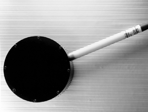
Model 6000-100 CT Dose Probe consists of a pencil-type ionization chamber with a sensitive length of 10cm and an active volume of 3.2cm³.
Model 6000-200 CT Dose Probe consists of a pencil-type ionization chamber with a sensitive length of 10cm and an active volume of 10cm³. This larger active volume greatly improves signal strength to over three times that of a typical 3cm³ CT probe. The outside diameter of the 6000-200 chamber is 12.7mm in diameter, requiring no adapter to fit into existing CT dose phantoms.
Both probes feature a 0.9 meter flexible, low-noise cable which is terminated in a male BNC coaxial connector for signal and a banana plug for bias. A triaxial BNC or TNC termination is available on request.
The 6000-100 and 6000-200 CT Dose Probes are designed specifically to read out on the Cardinal Health Model 8000 NERO®mAx X-ray beam analyzer, but with an appropriate connector or adapter, it can be used with any high-quality dosimetry electrometer. When the 6000-100 and 6000-200 are used with the Model 4000+ and 4000M+ X-ray beam analyzers, or the RAD-CHECK® PLUS, appropriate correction factors must be applied.
Model 007, CT Dose Phantom Set
The CT dose phantoms were designed in accordance with the Food and Drug Administration’s performance standards for diagnostic X-ray systems, which include regulations specifically applicable to CT systems (21CFR1020.33).
iii. Film QA
Hand-Held Digital Densitometer & Sensitometer
Model 331, Digital Clamshell Densitometer
Although compact and lightweight, the Model 331 Transmission Densitometer does not compromise on performance. For example, you get a 0-4.0D measuring range, accuracy of ±0.02D and repeatability of ±0.01D. With these rigid standards, along with the instrument’s rugged design, you will find that the Model 331 is ideal for both lab and field use.
A self-contained light table gives the densitometer added versatility — there is no need for an external light source. The light table is carefully designed for easy measurements on all areas of film up to 11 inches wide.
Other features include push button null, certified step wedge, low-battery indicator (typically after 2000 readings), battery eliminator (optional) for operation from an AC power source and an instrument carrying case.
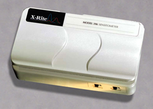
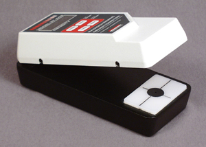
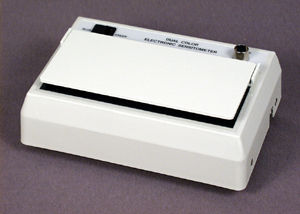
Model 396, Dual-color Sensitometer
Improved accuracy and repeatability are the hallmarks of the Model 396 Dual-color Sensitometer. It offers the latest technology with greatly improved blue and green film performance. It also features an exposure time control to adapt to conventional X-ray or cine film QC requirements.
Imagine the convenience of a sensitometer this small that exposes a 21-step wedge on green-or-blue-sensitive X-ray film, roll film or cine film. What’s more, it is simple to use. Just set the instrument to proper exposure for the film being used. Place the film inside the 396, then close and firmly press down the cover. When you hear the beep, the exposure is complete.
Other features include operation on a single 9-volt alkaline battery (with a life up to 10,000 exposures) and an on-off switch to prevent accidental battery drainage during transport.
The model 396 is ideal for mammography, mobile labs and cine film users.
Model 07-443
Hand-Held Densitometer
State-of-the-art features combined in a compact, handheld unit. Become a “speed reader" with this accurate, rugged portable clamshell densitometer. It has today’s most wanted features for go-anywhere quality control testing, from darkroom to darkroom, from lab to field use.
The Model 07-443 is easy to use: lift the shell and insert the test film; close the shell and press the READ button. The measured optical density appears on the 3-digit liquid crystal display. The self-contained light source makes it convenient to use anywhere. Includes a carrying/storage case and a five step Density Tablet.
Model 07-417
Dual-color Sensitometer
This compact, precision instrument is ideal for maintaining consistent, high-quality film processing. Also, processing conditions in multi-processor departments may be standardized. In the past, this was difficult in departments using varied film/ screen combinations in different areas. Now, proper exposure of either blue- or green-sensitive X-ray film is easily accomplished with no need for internal adjustments.
The Model 07-417 sensitometer features a 21-step density wedge with 0.15D increments. An innovative, dual-color, electroluminescent light source provides precisely-controlled, repeatable exposures. The desired color is selectable with a front-panel switch.
Model 07-424
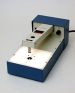
Digital Densitometer
This easy-to-use digital densitometer is a precision instrument that quickly measures the optical density of film. Constructed of rugged steel, compact enough to fit on any crowded worktable, it offers exceptional accuracy (±0.02 optical density). The optical density value is displayed in bright LED numerals on the detector arm. The sample table measures 6-1/8" x 10-1/2" with a 4-3/4" by 5-1/2" illuminated section. The large throat clearance will accommodate films up to 14" x 17" and provides ample room for positioning any selected area under the detector.
The detector is a hermetically-sealed silicon photodiode. The detector lamp is at full brilliance only during the actual measurement, thus preventing heating problems and assuring long lamp life with minimum wavelength shift or other degradation. The lamps are conveniently located for easy replacement when it becomes necessary to do so.
Model 07-440, Digital Densitometer with RS-232 Interface
With its optional RS-232 interface, the Model 07-440 allows you to automatically transfer data to a computer for storage and retrieval. By adding Film-Pro QC software, you can completely automate your daily processor QC program. Simply read each step on your 21-step sensitometer strip, send the data to your computer by pushing “send data" button, and let Film-Pro do the rest.
Model 07-460, Photographic Step Tablet
Allows standardization of radiographic film density. Consists of a strip of exposed, processed photographic film with 21 calibrated steps (0.05 to 3.05 density). Each step is 10mm x 35mm. The tablet is 250mm long x 35mm wide.
Model 301
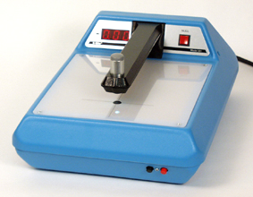
Digital Densitometer
Provides Accurate QA for Your Film Processor
The end product of your X-ray, microfilm or photographic processing equipment is only as good as the quality control techniques that you use. The Model 301 densitometer is designed to help you keep your film processor producing optimal results.
This densitometer will detect variations in film density far more subtle than can be seen with the naked eye. Its wide measurement range allows you to determine base fog and accurately determine density up to 5D.
A new optional feature has been added to the Model 301 – a serial RS-232 output. Now you can connect it to a serial input printer or an IBM-compatible computer. This provides a convenient record or an opportunity to use a computer to calculate the data from a step wedge.
Convenient Operation
The Model 301 is easy to use. Measurements are made on a push-and-read basis. The display consists of large LED numerals which can be read easily in bright or dim light. Internal memory and the null button allow the operator to make comparative density measurements across a piece of film.
Other quality features include a lighted work table, simple calibration procedure and rugged construction. The densitometer will remain in proper alignment even though it may be picked up by the arm.
The Model 301 is UL-listed and is standards-traceable through the NIST. This is probably the most widely-accepted and used densitometer for medical and industrial X-ray requirements and in the micrographic industry.
Model PQA-185
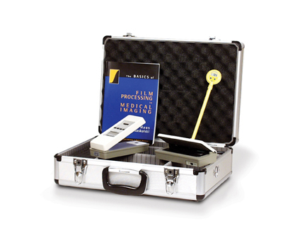
Processor QA Kit
Quality Assurance (QA) in radiology begins with the film processor. The processor is the single most influential source of problems in the diagnostic imaging department.
To test all the parameters of the processor, the PQA-185 Processor Quality Control Kit has been designed with all instruments contained in one storage/carrying case. Included in this kit is a portable blue/green sensitometer, portable handheld densitometer, a digital stainless steel stem thermometer and the text book “Basics of Film Processing in Medical Imaging".
With these tools, daily processor quality control can be completed conveniently and early detection of possible problems can be caught before they lead to poor quality images or expensive repairs to the processor.
Model DT-4339
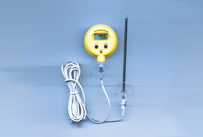
Digital Thermometer
Minor shifts in developer temperature can have a detrimental effect on the film contrast and density. In order to achieve and maintain appropriate film speed, film contrast and film fog levels, the developer temperature must be monitored on a regular basis. To maintain this temperature, a high-quality, accurate thermometer must be used, such as the DT-4339.
This thermometer is a perfect choice for all film processor quality control applications. It features a choice of Fahrenheit or Celsius units of temperature, maximum/minimum temperature memory and battery operation.
The thermometer is supplied with an NIST-traceable calibration certificate, suction cups and Velcro®. With this thermometer there is no chance of mercury contamination of your processor.
iv. Nuclear Medicine
Model CRC®-127R
Nuclear Medicine Dose Calibrator
The Model CRC®-127R now replaces the CRC®-7(R) and CRC®-12(R) by combining the best features of both of these units into a new package. The CRC®-127R continues the tradition of outstanding performance characteristics and a worldwide reputation for excellence for which Capintec dose calibrators are known.
By incorporating the speed of the CRC®-7 and the Becquerel/Curie conversion plus auto-ranging functions of the CRC®-12, the CRC®-127R provides simplicity of operation to meet your needs.
The CRC®-127R provides eight preset calibration settings for the most commonly used radionuclides: Tc-99m, Tl-201, In-111, Ga-67, I-131, I-123, Xe-133 and Moly assay. The CRC®-127R allows calibration settings for more than 200 other nuclides. Precision potentiometer dials in calibration settings for any radionuclide. The bias battery used in the CRC®-127R offers stability when the current in your lab does not. The battery has a nominal 10 year life.
The CRC®-127R is the lowest-priced dose calibrator from Capintec and offers the longest warranty of any dose calibrator sold.
Model CRC®-15R
Nuclear Medicine Dose Calibrator
The Model CRC®-15R is Capintec’s most popular dose calibrator, and for a good reason. Taking advantage of the latest technology, the CRC®-15R is an autoranging dose calibrator, giving you added speed, accuracy and easy-to-use features. There are nine preset nuclide keys plus five user-defined keys that may be preset to any nuclide you require. The advanced system also allows selection of over 86 different nuclides through the alphanumeric keys by simply entering the nuclide name and number and pressing ENTER.
For generator users, the CRC®-15R automatically calculates the Moly-99/Tc-99m ratio. An optional printer provides syringe/vial peel-off labels that show Tc-99m activity, Moly-99/Tc-99m ratio and total molybdenum content. Full-size pages are printed of QC tests to satisfy state and NRC regulations.
For unit dose users, the speed of the CRC®-15R reduces time spent waiting for the final check before patient administration. The large backlit screen always identifies which nuclide is being counted. No more guessing which nuclide button is activated.
CRC®-15R is also the smallest available dose calibrator. Its screen design allows viewing with the unit placed vertically on the shelf or wall-mounted. With the easy TEST section, you are stepped through automatic background and zero adjustments as well as accuracy, constancy and system testing.
v. Phantoms
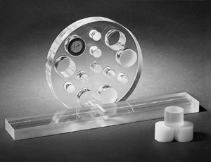
Model 18-220
Mammographic Accreditation Phantom
Radiology professionals and patients rely on mammographic systems to produce breast images of the highest quality. However, in sensitive mammographic imaging equipment, small deviations in system performance can lead to substantial decreases in image quality.
Without the phantom to serve as a baseline test, even the most experienced professional can miss the subtle degradation of image quality that occurs gradually over time in sensitive mammographic equipment. Weekly checks with this phantom can alert you to minor deviations in image quality before they impair the diagnostic accuracy of patient films.
The Model 18-220 Mammographic Accreditation Phantom is designed to attenuate the X-ray beam in the same way as a human breast compressed to 4.0 to 4.5cm. Test objects of different sizes and shapes are embedded in a wax block enclosed in an acrylic base. These test objects represent malignancies or small breast structures: simulated microcalcifications, fine nylon threads for fibrils and ductal structures, and parts of spheres for tumor-like masses.
When the Mammographic Accreditation Phantom is used to create an image, the mammographic imaging system should detect a minimum of 4 fibrils, 3 groups of specks and 3 masses in the phantom. If the phantom image is good, the system may need no further testing. However, if the objects in the image cannot be seen, or if the visibility of these objects decreases over a period of weeks, it will be an indication that the system needs additional testing. Variables to check include the film processor, kVp, phototimer accuracy, film/screen contact and radiation beam quality.
The Mammographic Accreditation Phantom will assist in complying with the American College of Radiology (ACR) Accreditation Program requirements. Since this phantom adheres to ACR phantom specifications, it will assist you in meeting the requirements of MQSA.
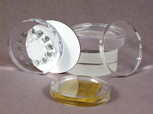
Model 303ACR, Chamber Holder
This acrylic chamber holder is designed to conveniently position the Model 303 mammographic ionization chamber close to the Mammographic Accreditation Phantom, with the center of the chamber’s sensitive volume flush with the top of the phantom, for simultaneous in-air dose measurement and artifact imaging. Its wide base maintains parallelism to the pressure plate, and the ionization chamber is held securely in place by a nylon screw. The backscatter is negligible due to its hollow construction.
Model 96035ACR, Chamber Holder
Identical to 303ACR chamber holder, except that it is designed to hold a Keithley Model 96035 and CNMC Model 315 15cc diagnostic ion chambers.
Stereotactic Biopsy Phantoms
With the increasing use of stereotactic breast biopsy procedures, it is essential that radiology healthcare providers maintain and increase their needle biopsy skills.
A comprehensive mammography quality control program must provide assurances that all aspects of the mammography equipment are operating at optimum levels. The automated stereotactic breast biopsy procedure depends on several variables for accurate needle placement. Thus, for patient safety, this system must be properly maintained and evaluated.
These phantoms were designed to assist in the training of technologists and physicians in the use of a stereotactic system, and for verifying the proper operation of mammographic stereotactic biopsy systems.
Both phantoms closely mimic properties of the human breast. They are ideal teaching tools and practice mediums for mammographic needle biopsy procedures. They are also excellent quality assurance testing devices for stereotactic systems, and should be used whenever a new system is installed or repaired, to insure accurate needle placement.
The phantoms should be stored in a cool dry place and discarded after all the tumors have been aspirated.
Model 18-250
Digital Mammography Evaluation Phantom
Until now, there has not been an easy way to compare the image quality of conventional and digital biopsy mammography units. The Model 18-250 Digital Mammography Evaluation phantom allows you to do just that. The field of view on the digital system is typically much smaller than the 24 x 30cm field of view on the conventional mammography units.
In order to image the Mammographic Accreditation Phantom specified by ACR on the biopsy units, the user had to move the phantom to various positions, overlapping to be sure that all objects were imaged. This is a very inconvenient, time consuming task.
The small size of the Digital Mammography Evaluation Phantom enables you to image the unit entirely in a single exposure. The objects are some of the same found in the Mammographic Accreditation Phantom specified by the ACR, so it makes comparison of the two imaging systems easily.
Model 18-251
Contrast & Resolution Mammo Phantom
On digital units, this phantom can test the high contrast spatial resolution of the system with the results being viewed on the monitor. The focal spot high contrast resolution can also be determined by placing a conventional mammography cassette behind the phantom and making an appropriate exposure. The greyscale step wedge can be used to visually test the dynamic range of the digital imaging system by viewing it on the monitor. It can also aid in the setup of hardcopy cameras.
It is now being suggested that a resolution test pattern from 5-20 LP/mm be used to evaluate the condition of the focal spot. Instead of making focal spot measurements which can be ambiguous, an accurate determination of the X-ray tube’s resolution ability can be measured by using this test pattern.
On conventional mammography units, the phantom can be used to meet the ACR guidelines for testing focal spot resolution. The ACR suggests placing a resolution target 4.5cm above the image receptor and imaging twice, once parallel to the anode-cathode axis and once rotated 90 degrees. With two resolution targets, this can be achieved in a single exposure. The greyscale step wedge can also be used to check the dynamic range of the entire system, indicate processing problems and variations in film emulsion. The phantom is designed with an extended top edge to aid in positioning on recumbent biopsy tables.
When ordered with the phantom, the test patterns will be inserted into the phantom and shipped complete. If you already have the 07-555 pattern, you may quickly and easily insert the pattern by removing two screws on the top of the phantom. Be sure to place the 07-555 into the phantom block in either one of the orientations shown in the photo.
The phantom allows a determination if the film appears similar to the soft copy display. For the best calibration possible, SMPTE Test Pattern Generator Model 07-495 should be used in addition to 07-555. Multi-format Medical Imaging Test Pattern Generator Model 07-499 is also available to meet this requirement.
Model 76-430
Mini CT QC Phantom
The Model 76-430 is a highly-versatile phantom designed for routine monitoring of the consistency of all the major parameters of CT image quality and radiation dose. Its unique, compact design allows for unparalleled portability, easy setup and reliable parameter determinations. It is perfect for use by physicists, technologists and service engineers.
The disc section consists of a 1" thick acrylic disc with a 6" diameter. Five of the six large holes are for the placement of inserts for the evaluation of CT number consistency. The sixth hole is for a high-contrast resolution insert or an optional low-contrast resolution insert. The seven small holes are for inserting an ion chamber at differing locations within the phantom. Six acrylic inserts are provided to fill these holes, leaving one hole to accommodate an ion chamber during a given measurement.
The disc section is attached to a rectangular acrylic bar containing a thin copper wire embedded along a central grove. This section of the phantom is used to evaluate laser beam alignment and accuracy of the slice thickness, slice spacing, slice contiguity and pilot scan to transverse, or longitudinal scan correspondence. This is achieved by exposing a non-screen film placed underneath the phantom and making several cuts while the phantom is advanced along the gantry in a pre-programmed manner.
Model 76-432
CT Spiral Phantom
The accurate indexing capability and exceptional image quality of computed tomography (CT) scanners not only guarantee the object’s location, size and shape, but also improve the diagnosis accuracy. The index and performance parameters of CT scanners cannot be confirmed without objects of known specifications. The Model 76-432 CT Spiral Phantom provides specific details necessary to confirm the integrity of both conventional and spiral scanning. What makes the phantom unique is that it allows the user to visually evaluate all test results in their image displays.
The phantom consists of five acrylic plates of different sizes, all affixed to a flat rectangular base. Specific hole patterns are drilled on each side of these plates. When imaging, the holes within the X-ray field will appear in the phantom images. By the appearance of the holes, both index and performance parameters can be confirmed qualitatively and quantitatively.
PIXY®
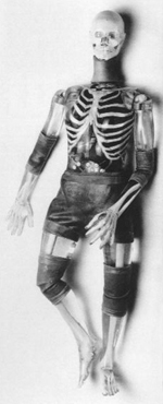
Anthromorphic Training/Teaching Phantom
PIXY® is used to demonstrate anatomy and evaluate positioning and imaging techniques, including kVp, mAs, contrast, optical density, OFD and TFD. Radiographs of PIXY® are optically equivalent in density and contrast to human patients. PIXY® permits unlimited exposures and tolerates trainee errors.
Anatomy – PIXY® shoulders have ball and socket joints; elbows and knees flex 90° and 100°. Hips flex 130° with 30° hyperextension.
A “frog" position is made possible by lateral flexion at the hips. The right hand is molded with fingers positioned for an AP view, while the left hand is in an oblique-lateral position. The left foot is in full plantarflexion; the right foot is in neutral position.
Neck vertebrae C1, C2, C6 and C7 were converted to mechanical nylon joints because the educators in the field prefer full positioning capabilities for the head. The design permits the remaining neck vertebrae to be fixed in a normal position, while assuring a full range of head motion.
PIXY® contains abdominal and pelvic organs: stomach, gallbladder, urinary bladder, kidneys, rectum and sigmoid flexure. These are air-filled, but accept water or contrast media and can be easily flushed after use. Custom fractures and custom pathologies are optional.
Materials – Highly-detailed polymer skeletons which reproduce shape, mass density and attenuation co-efficients of the cortical bone and spongiosa, allow continuous production of phantoms, instead of sporadic production due to limited availability, variable size and uncertain chemical composition of human skeletons. Nevertheless, human skeletons are available for users who desire them. There is a surcharge to cover the high cost of scarce natural skeletons and for added labor needed to rework them to fit PIXY®molds.
The matching of skeletons and soft tissues permits external and bony landmarks to coincide. The position of bones within the soft tissues is anatomically correct.
The detail cast into skeletons is considered a triumph of sculptural moldmaker’s craft. The skull, for example, has frontal and sphenoidal sinuses, ethmoidal and mastoid air cells and the auditory ossicle. Bone structures are radiographically visible.
Soft Tissues – PIXY® is available in opaque or transparent tissue equivalent materials. The transparent PIXY® has visible organs and skeleton except at the hips, knees, and elbows, which are opaque because, as on opaque PIXY®, latex coverings are needed to retain tissue-equivalent gels for soft-tissue continuity at these articulations. Two-ply coverings protect against gel leakage.
Standard PIXY® lungs are molded of tissue-equivalent foam with a mass density of inflated human lungs (0.30g/cc). They are connected to the oronasal cavity by the stem bronchi and trachea. The oro-nasal pharynx is filled with a nearly air-equivalent foam.
Optional animal lungs, which duplicate the intricate detail of the vascular trees, are available. These lungs are fixed in the inflated state and molded to conform to the pleural cavities of the phantom.
The pulmonary arteries are injected with the blood-equivalent plastic. In addition, low-, medium-, and high-contrast material may be selected by the user.
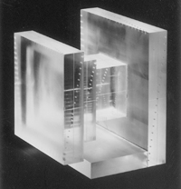
Sectional Phantoms
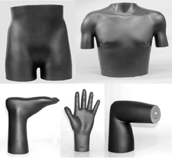
Anthromorphic Training/Teaching Phantom
Sectional phantoms are used when you require a human subject to train radiographers in the use of radiography equipment. The complete sectional phantom set and the individual sectional phantoms may be used repeatedly to learn how various positions and techniques will produce a variety of radiographic images.
A natural human skeleton is first reconstructed into the sectional molds of an average-sized male patient. With the bones set in place, the mold is filled with a specially formulated urethane material, the same as is utilized in the RANDO® phantom, which simulates the density of tissue and organs. This opaque material duplicates the X-ray absorbency, atomic number, and specific gravity of soft human tissue. Because the RANDO® material is virtually indestructible, these sectional phantoms will withstand substantial impact and continuous handling without damage.
Only natural skeletons are used in these anthropomorphic phantoms. Many skeletons reflect natural human characteristics such as lack of symmetry and distorted joints. As the technicians reconstruct the skeleton, minor adjustments may be made to facilitate positioning within the mold. The natural characteristics and the adjustments should not present any problems in the use of the phantom.
Model SK150, Head
The skull and cervical vertebrae are cast into the head phantom with an internal air cavity to represent the oral, pharynx and trachea anatomy. It is also available without the cervical vertebrae.
Model SK200, Thorax
The thorax phantom contains the chest and shoulder skeletal structures including the upper third of the humerus. The lungs’ lower density is duplicated with a special material, the same material as used in the RANDO® phantom lungs, which has an atomic number and specific gravity of human lungs at a median respiratory state. This material is formed to the contours of the rib cage.
Model SK250, Lower Torso
The lumbar vertebrae, pelvis and upper third of the femur are cast into the lower torso phantom. A hollow cavity reproduces the interior of the sigmoid flexure with the diverticulum and rectum. This cavity is connected to the surface by a duct that is sealed with an O-ring screw cap. The cavity may be filled with contrast media. It is not discernible when filled with water.
Models XA235-L & XA235-R, Left and Right Elbows
The elbow phantoms contain the lower third of the humerus and the upper third of the radius and ulna. The right elbow has a 90° flexion and the left elbow is extended.
Models XA231-L & XA231-R, Left and Right Hands
The hand phantoms contain the lower third of the radius and ulna along with the carpal and finger bones. The left hand is pronated and the right hand is relaxed.
Models XA245-L & XA245-R, Left and Right Knees
The knee phantoms contain the lower third of the femur, the upper third of the fibula and tibia, and the patella. The right knee has a 90° flexion and the left knee is extended.
Models XA241-L & XA241-R, Left and Right Feet
The foot phantoms contain the lower third of the fibula and tibia along with the tarsal and toe bones. The left foot is relaxed and the right foot is in plantar position.
Model SP500, Complete Sectional Phantom
The complete set consists of 11 sectional phantoms including head, thorax, lower torso and the left and right elbows, hands, knees and feet. All come packed in a convenient wooden storage case.
RANDO® is a registered trademark of the Phantom Library
v. Test Tools
Mammo Phototimer
Consistency Test Tools
A mammographic unit’s Automatic Exposure Control should be capable of maintaining optical density within ±0.15 OD as the voltage is varied from 25 to 35 kVp, and as breast thickness is varied from 2 to 8 cm for each technique. Test images taken of uniform phantoms of varying thicknesses should not differ by more than ±0.30 OD from each other. These tests should be carried out over the kVp range customarily used by the mammography center.
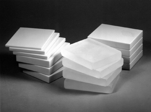
The phototimer consistency test tools are available in two materials: acrylic and, for more accurate results, breast tissue equivalent BR-12 material.
BR-12 is a designation (D.R. White et al.) of certain epoxy resin formulations that react to X-rays in the mammographic energy range (15-30 keV) in the same manner as the human tissue. The tissue simulation properties for these slabs are maximized at 20 keV (28kVp±). The glandular equivalency of this material is 45% in the mammographic range.
X-Ray Test Tools 1
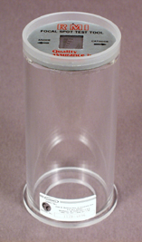
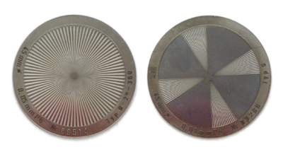
112B Focal Spot Test Tool
The focal spot size of an X-ray tube is of crucial importance in determining the detail of an X-ray image. The Focal Spot Test Tool was developed to allow easy and accurate interpretation of the effective focal spot size of radiographic X-ray tubes. The Model 112B consists of a metal target with twelve bar pattern groups. Each group consists of six slots with three slots perpendicular to the other three. The sizes and spacing of the slots in the 12 groups decrease by steps of 16% from 0.84 LP/mm to 5.66 LP/mm. The test pattern is mounted in the center of an acrylic disc 7.6cm in diameter that contains a lead shield. The Model 112B is easier to interpret than the pinhole image or a star pattern for effective focal spot measurements.
117 Radiographic Aluminum Step Wedge
This step wedge is constructed of homogeneous, high-purity aluminum and is designed to provide incremental exposures to X-ray film by the increased aluminum thicknesses in each step. The Model 117 provides a useful means of comparing the characteristic curve of various film-screen combinations, mAs reciprocity, and, if done very carefully, sensitometry. For sensitometry totally independent of X-ray generator variations, use a dedicated sensitometer/densitometer.
118 Mammographic Aluminum Step Wedge
Constructed of homogeneous, high purity aluminum, this step wedge provides incremental exposures to mammographic X-ray film by the increased aluminum thicknesses in each step. The step wedge provides a useful means of comparing the characteristic curve of various film-screen combinations, mAs reciprocity and, when carefully performed, sensitometry. For sensitometry totally independent of X-ray generator variations, use a dedicated sensitometer/densitometer.
X-Ray Test Tools 2
132 Tomographic Test Tool
Designed to test the imaging capabilities of the tomographic system, the Model 132 can be used to:
- Determine the location of the cut plane.
- Determine the thickness of the cut.
- Test the overall resolution of the cut plane.
- Test the X-ray exposure uniformity.
- Determine the path of the beam during exposure, for both linear and multi-directional units.
141 & 141H High-Contrast Resolution Test Tools
One important measure of your fluoroscopy system is its high contrast resolution. This test can assess the resolving power of your system and can be accomplished easily with Models 141 and 141H.
Both high-contrast resolution test tools consist of eight patterns of copper wire mesh in a pie shape. Each is labeled with lead numbers for easy visualization. Model 141 is used for standard radiographic systems with resolutions between 16 and 60 mesh, and Model 141H is used for systems with higher resolution such as those used in cardiology suites, where resolution is between 60 and 150 mesh.
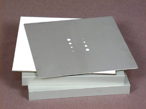
142D & 143D Film/Screen Contact Test Tools
Inspection of cassettes for good film/screen contact and screen integrity is an important but often overlooked quality control procedure. Poor film/screen contact can be the reason for areas of increased density, reduced density and blurring.
X-Ray Test Tools 3
151 Low-Resolution Test Tool
An Automatic Brightness Control (ABC) for fluoroscopy compensates for variation in patient thickness, X-ray field size, image intensifier magnification modes and other variations of the system. The Model 151 Low-Contrast Resolution Test Tool evaluates the system’s ability to compensate for these variations while maintaining good contrast and detail.
The Model 151 consists of two aluminum blocks, one lead blocker and an aluminum resolution plate with two sets of decreasing diameter holes. Other tests performed are table top exposure rate in R/min for fluoro units and the phototimer performance of spot film devices.
144 Grid Alignment Test Tool
The Model 144 is designed to test proper grid alignment with respect to the central ray of the X-ray tube. Grid misalignments, such as lateral decentering or tilting of the grid, are not easily recognized and can have an effect on image contrast as well as increasing patient dose. These types of misalignments can only be detected through regular quality control testing with the Model 144.
161B Collimator Test Tool & 162A Beam Alignment Test Tool
The Model 161B is designed to evaluate the collimator light field and X-ray field congruence. It is constructed of brass so that centimeter etchings on its surface can give a direct ruled dimension on the radiograph. It is calibrated to show misalignments to within 0.5 cm.
The Model 162A provides a simple test of the X-ray beam’s alignment. When used with the Model 161B, misalignments of 1% and 2% can be visualized. It is constructed as a plastic cylinder with one steel ball at each end. If everything is in alignment, the steel balls will be superimposed on the radiograph.
X-Ray Test Tools 4
1151 Radiographic Contrast/Detail Test Tool
A useful method of assessing the overall image quality of a fluoroscopy system is by defining its ability to detect small objects with small differences in contrast from the background.
The Model 1151 consists of an aluminum plate with a 10 x 10 matrix of holes that vary in diameter and depth. For a hole diameter, the depth of the hole which can just be visualized is defined as the contrast for that diameter. A contrast detail curve of the fluoroscopic system can be established by plotting the diameter of the hole vs the depth of the hole that is being visualized.
AFS-1 Aluminum HVL Attenuator Set
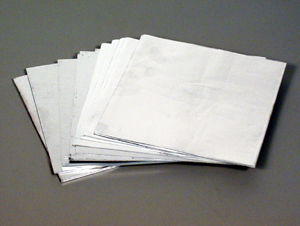
Consists of sixteen 10 x 10cm absorbers of various thicknesses; six of 1mm, two of 0.5mm, four of 0.1mm and four of 0.05mm made of 1100 aluminum with a purity of 99%. Total thickness of the set is 7.6mm.
07-434 High-Purity Aluminum HVL Attenuator Set
When doing HVL measurements with a mammography unit, it is recommended that highest-purity aluminum be used. The set consists of five high purity (99.99%) aluminum filter, 10 x 10cm, 0.1mm thick, which permit more accurate half-value layer determination on mammography machines.
07-431 Copper HVL Attenuator Set
For HVL determination of high-range X-ray generators (140 to 400 kVp). Set consists of ten 10 x 10cm absorbers, including four of 1mm, two of 0.5mm, and four of 0.1mm, for a total thickness of 5.4mm.
X-Ray Test Patterns
Star Test Patterns
Focal spot size can be determined by observing the regions of blurring which occur when the pattern is radiographed by an X-ray source of finite dimensions. Radiation from different areas of the focal spot will cause a periodic blurring of the pattern due to penumbra effects. Knowledge of the geometric factors and the distance from the center of the pattern to the region where blurring occurs will permit the calculation of the focal spot size.
The star patterns listed below are handmade, ultraprecision types.
07-555 Resolution X-Ray Test Pattern
Resolution X-Ray Test Pattern is specifically designed for evaluation of focal spot performance protocol in Mammography.
It is now being suggested that a resolution test pattern from 5-20 LP/mm be used to evaluate the condition of the focal spot. Instead of making focal spot measurements that can be ambiguous, an accurate determination of the X-ray tube’s resolution ability can be measured by using this test pattern. The Model 07-555 is manufactured from 0.02mm thick gold foil, 25mm long and 10mm wide. The pattern has 13 segments, from 5 LP/mm to 20 LP/mm. Radio-opaque numbers indicate the resolution (in LP/mm) of each group.
MC-1 Pocket Optical Comparator
Ideal for checking small parts, linear measurements, hole diameters, thread sizes and slit camera images. Unlike lower priced comparators with only a simple lens, the MC-1 incorporates a triplet lens. It provides an extremely flat field over the entire reticle area. The clear, acrylic cell allows the outside light to illuminate the subject. The comparator reticle is different from ordinary reticles in that the fine ink-filled markings are on the outside of the reticle. This is done so that the reticle scales are always in direct contact with the object and you always get optimum focus, freedom from parallax and accurate measurements. The scale reticle is optical glass with a pigment-filled pattern, 20mm long with 0.1mm per division.
Model 181-4000
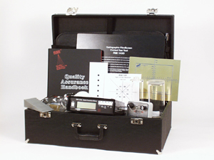
Radiographic/Fluoroscopic QA Kit
The Radiographic/Fluoro Kit contains the necessary test instruments for doing routine quality control tests of radiographic, fluoroscopic and tomography X-ray units. Each test tool within the kit is designed to evaluate one of the many important imaging parameters within the X-ray system.
Instructions for all the tools are in a hardcover binder for easy location and availability. The instructions are “cookbook" easy so that personnel will find the procedures easy to do and understand. The user will be able to immediately begin recording data on the sample quality control forms that are included with the instructions.
Model 120-20 Visi-X
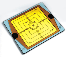
X-Ray/Light Field Alignment Tool
The Model 120-20 Visi-X is a new concept in Quality Control and Service. It is a cassette-shaped instrument for checking the light and radiation field coincidence on x-ray equipment. The Visi-X can also be used for checking the centering of the Bucky tray.
The Visi-X is constructed of acrylic and its operation is based on the long persistence afterglow principle of its phosphor screen. The phosphor is non-radioactive and is confined by acrylic plates. A daylight filter protects the phosphor from unwanted excitation from light sources.
To use, simply darken the X-ray room and place the Visi-X under the X-ray tube. Adjust the light field and make the exposure. The radiation field will immediately be visualized by the glow of the special phosphor compound. This afterglow will last for several minutes. Misalignments as little as ±0.5mm will be clearly shown on the built-in scale. No film is needed, therefore no time is lost going back and forth to the film developer.
When viewing the ordinary intensifying screens you will expose yourself to unnecessary radiation. With the Visi-X, you do not have to be present in the X-ray room, therefore there is no radiation hazard.
The Visi-X will conveniently check the light and radiation field coincidence on your conventional diagnostic, dental and mammographic X-ray equipment. A magnetic lock allows Visi-X to be used in vertical and upside-down positions.
vi. Ultrasound
Ultrasound Phantoms
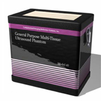
Model 040 General Purpose Multi-Tissue Ultrasound Phantom
The Model 040 offers a reliable medium which contains specific, known test objects for repeatable qualitative assessment of ultrasound scanner performance over time.
This phantom is constructed from the patented, solid elastic material, Zerdine®. Zerdine® is not affected by changes in temperature. Zerdine® allows more pressure to be applied to the scanning surface without subsequent damage to the material. At normal room temperature, Zerdine® will accurately simulate the ultrasound characteristics found in human liver tissue. The Phantom includes detachable scanning wells to accommodate large sector probes and endocavity probes and a rugged carrying case.
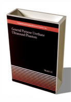
Model 042 General Purpose Urethane Ultrasound Phantom
The Model 042 enables repeatable, qualitative assessment of ultrasound scanner performance over time.
The Model 042 Axial Resolution Target is constructed from a proprietary urethane matrix, housed within a rigid PVC container with three separate scanning windows. It allows for depth of penetration, uniformity, distance calibration, resolution and lesion detectability assessment. The Model 042 is sold with a three-year warranty. This phantom includes an in-house certification traceable to NIST, user’s manual, ultrasound physics textbook and a carrying case.
X-Ray Equipment QA
DIDO2000 Series
The DIDO2000 Series diagnostic dosemeters cover all fields of X-ray applications. No matter if conventional or digital modality, the meters can be used for measurements in Radiography, (Pulsed) Fluoroscopy, DSA, Dental, 3D (CBCT), and Mammography.
Nonius
The Nonius is an easy-to-use but very sophisticated measuring instrument to verify size and quality of X-ray fields. It can also be used to analyse the properties of fanned X-ray beams.