


Dosimetry Phantoms
Please click the product you want to learn more about:
Enquiry Now
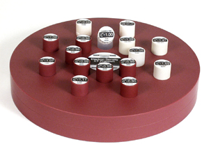
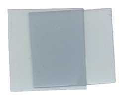
Build-up Caps
Build-up Caps
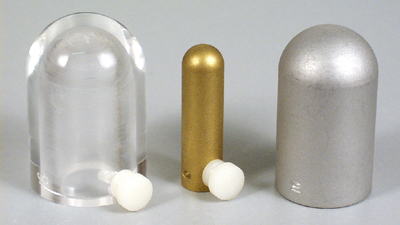
The total build-up should be calculated using the wall thicknesses and thimble thickness with the appropriate density. For detailed statistics on the thicknesses available, please open the specification sheet.
Electron Density
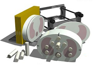
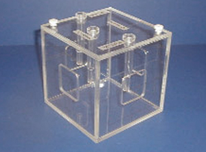
Model EDP-62
Electron Density CT Phantom
The accuracy of radiation oncology treatment planning systems is heavily dependent upon precise CT analysis of the patient anatomy which is to be irradiated. Physicists performing treatment planning need accurate tools to evaluate CT scan data, correct for inhomogeneities, and to document the relationship between CT number and tissue electron density. The Model EDP-62 Electron Density CT Phantom was designed and developed specifically to meet this requirement.
The EDP-62 phantom can be configured to simulate the head or abdomen. Tissue-equivalent plugs made of various tissue- and water-simulating materials can be positioned at 17 different locations within the scan field. Special marker plugs check the CT scanner’s distance measurement accuracy.
Scanning this phantom on a periodic basis provides data useful for the Quality Assurance program of both the CT scanner and the treatment planning system. A padded carrying case is included.
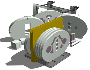
Model 467
Electron Density CT Phantom
Accurate corrections for tissue inhomogeneities are a critical part of isodose treatment planning. Although most computerized treatment planning systems currently use CT image data, these systems frequently use empirical formulae in computing correction factors for tissue inhomogeneities. The Model 467 Electron Density CT Phantom can be used to calibrate the CT unit by establishing the relationship between the electron density of various tissues and their corresponding CT number (in Hounsfield Units, HU). This data can then be transferred to the computerized treatment planning system for more accurate corrections for tissue inhomogeneities.
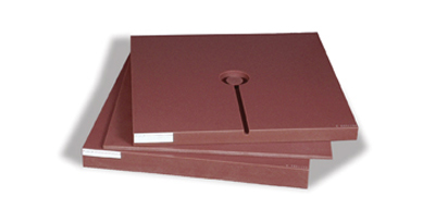
The Electron Density CT Phantom consists of a Solid Water disc approximating the size of an average pelvis. A matrix of sixteen holes in the disc hold interchangeable plugs made of various tissue and water-simulating materials. The physical density (g/cm³) and electron density relative to water of the plug materials are listed in the user’s manual. The phantom also has a pattern of small air holes with known spacings for checking the CT scanner’s distance measurement accuracy. A carrying case is included.
Scanning this phantom on a periodical basis provides data useful for the Quality Assurance Program of both the CT scanner and the treatment planning system.
IMRT
Model IMRT-2H5
IMRT Homogeneous Phantom
The Model IMRT-2H5 homogeneous phantom is designed to address the complex issues surrounding the commissioning and comparison of treatment planning systems while providing a simple yet reliable method for verification of individual patient treatment plans and delivery.
The phantom is homogeneous and elliptical in shape. It represents human anatomy in size and proportion. Measuring 30cm x 30cm x 20cm thick, the phantom is manufactured from unique proprietary materials that faithfully mimic water within 0.5% from 25keV to 50MeV.
Water-equivalent, interchangeable rod inserts for ion chambers allow for point dose measurements in multiple planes in the phantom and film calibration. The phantom also supports film dosimetry with not only standard radiographic films but also Gafchromic media at mid-plane in the phantom for analysis of dose distributions. Optional inserts are available to support a variety of other detectors including TLDs, MOSFETs and diodes.
The surfaces of the phantom are etched for ease of laser alignment, and CT markers ensure accurate film to plan registration. An alignment plate and compression device are included.
Model IMRT-2H9K
Point Dose Measurement Phantom
With the IMRT-2H9K phantom, you may choose any point dose location within a circular area with a diameter of 11.2cm by simply rotating a cylinder within a cylinder etched with indices for precise alignment. Each cylinder accepts an interchangeable rod that can be drilled for a variety of ion chambers, diodes, MOSFETs or TLDs. One water-equivalent and one bone-equivalent rod drilled for a specified detector are included. Additional lung-and bone-equivalent rods can be positioned at any location within the circular area for assessment of heterogeneity correction.
The phantom is homogeneous, made from proprietary material that faithfully mimics water within 0.5% from 50KeV to 25MeV. This unique material eliminates the need for correction factors, thus improving accuracy and saving time. The phantom simulates the patient through the entire process from CT data acquisition and planning to delivery and dose verification.
The shape is elliptical and approximates the size of an average patient. Tissue-equivalent, interchangeable rod inserts for ion chambers allow for point dose measurements in multiple planes in the phantom and film calibration. The phantom also supports film dosimetry with not only standard radiographic films but also Gafchromic media. CT to film markers ensure accurate film to plan registration. Close placement of detectors to film improves film calibration. Optional inserts are available to support a variety of other detectors including TLDs, MOSFETs and diodes.
The center cylinder may be removed to simulate head and neck setups.
When fully assembled, the phantom measures 30 x 20 x 30cm. An alignment plate and compression device are included.
Model IMRT-2HN
IMRT Head & Neck Phantom
The Model IMRT-2HN head and neck phantom is manufactured from water-equivalent plastic material. It faithfully mimics actual tissue within 1% from 50keV to 25MeV for accurate simulation from CT planning to treatment delivery.
A unique interchangeable plug design and cross sectioning allow various phantom versions to accommodate a multitude of dose measurement devices including ion chambers, film and gels. The plug locations begin in the center and 3, 4, 5 and 6cm from center, 90 degrees apart. Cross sections accommodate standard ready-pack films and special fiducial markers to allow for easy CT-to-film registration. A 6.35cm thick section is machined to accommodate a film stack for 3D image reconstruction or a Barrex cylinder for dosimetry gel.
The phantom is round in shape with a diameter of 16cm, and 30cm long when fully assembled. The IMRT-2HN includes an alignment plate with two rails that positions the phantom sections and a holding device that compresses the sections and films during irradiation. Both devices are made from relatively water-equivalent plastics to avoid any influence on the measurement results.
Model IMRT-2LFC
IMRT Thorax Phantom
The Model IMRT-2LFC thorax phantom is designed to address the complex issues surrounding the commissioning and comparison of treatment planning systems while providing a simple yet reliable method for verification of an individual patient’s treatment plans and delivery.
The phantom is elliptical in shape and represents an average human torso in proportion, density and structure. Measuring 30cm x 30cm x 20cm thick, the phantom is manufactured from unique proprietary materials that faithfully mimic water within 1% from 50keV to 25MeV.
Tissue-equivalent, interchangeable rod inserts for ion chambers allow for point dose measurements in multiple planes within the phantom. Hole placement allows for verification in the most critical areas of the chest. One half of the phantom is divided into 12 sections, each section is 1cm thick, to support film dosimetry with not only standard radiographic films but also Gafchromic film. Optional inserts are available to support a variety of other detectors including TLDs, MOSFETs and diodes.
The surfaces of the phantom are etched for ease of laser alignment. Optional CT markers are available to ensure accurate film to plan registration. An alignment plate and compression device are included.
Model IMRT-2PRA
IMRT Pelvic 3D Phantom
The Model IMRT-2PRA 3D pelvic phantom is designed to address the complex issues surrounding the commissioning and comparison of treatment planning systems and verification of individual patient’s treatment plans and delivery.
The phantom properly represents human pelvis anatomy in shape, proportion and structure as well as density. This enables thorough analysis of both the imaging and dosimetry system. The phantom is manufactured from unique proprietary materials that faithfully mimic bone and water within 1% from 50keV to 25MeV.
The phantom is elliptical in shape, approximates the size of an average patient and has a tissue-equivalent three dimensional skeleton. Tissue-equivalent, interchangeable rod inserts for ion chambers allow for point dose measurements in multiple planes in the phantom and film calibration. The phantom also supports film dosimetry with not only standard radiographic films but also Gafchromic media. Optional inserts are available to support a variety of other detectors including TLDs, MOSFETs and diodes.
A 6.35cm thick section is machined to accommodate a film stack for 3D image reconstruction or a Barrex cylinder for dosimetry gel. With the aid of three spacers, the film stack or the gel cube can be positioned in six different locations within the section.
Included are four different electron density reference plugs which can be interchanged in five separate locations within the phantom. The surface of the phantom is etched with grooves to ensure proper orientation of the CT slices and accurate film-to-plan registration.
When fully assembled, the phantom measures 30 x 20 x 21.4cm. An alignment plate and compression device are included.
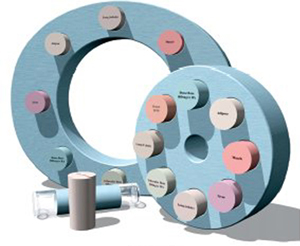
ISIS QA-1
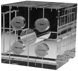
Geometric QA Phantom
The ISIS QA-1 Phantom was designed to provide an easy, low-cost approach to the daily, monthly and annual QA tasks for the Physicist and Therapists. The ISIS QA-1 phantom will aid in verifying the geometric laser position accuracies with multiple laser systems within your department. The ISIS QA-1 also provides the Physicist and Dosimetrist the ability to verify electron beam density values produced by your CT / CT-Simulator. Staff members scan the four unique density value inserts, then transfer this image to the RTP system for verification of the electron density values of the Bone, Water, Inhale and Exhale Lung density inserts. Comparing the individual value for each known density value, the user can quickly verify CT image electron density values for treatment planning image QA.
Additionally, the ISIS QA-1 provides an internal known object insert that is scanned with the CT / CT-Simulator. With this multiple image slice set you can create a Treatment Plan / Virtual Simulation plan of the known object for size and location verification though your RTP and Virtual Simulation system. The ISIS QA-1 then goes one step further to use these known geometric phantom positions for verification of the laser positions as verified with the scanned ISIS QA-1 phantom. This QA process provides a geometric QA of the processed RT Plan for use with IMRT treatment machine lasers and mechanical treatment field setup verifications. The dose chamber insert will provide the physicists the ability to quickly measure single-point expected dose values without using additional phantom devices. This removable insert is matched to your existing standard dose chamber (model and manufacturer of chamber must be specified).
The ISIS QA-1 helps standardize the QA program for the installation engineer, maintenance engineer, therapist, dosimetrist and/or physicist. These QA tasks are accomplished by using a common single QA alignment/ verification tool, thus producing a common geometric theme within the radiation therapy department prior to implementation of an IMRT or standard external beam treatment program. The continuing review of the geometric accuracy between all department systems is important with using today’s high-quality and technical treatment protocols.
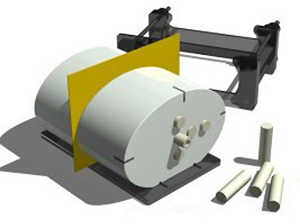
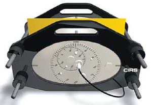
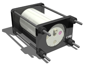
Model DTP-008
Dynamic Thorax 4D QA Phantom
The Model DTP-008 Dynamic Thorax Phantom is designed to investigate and minimize the impact of organ motion and patient positioning errors in radiation therapy. It is the first commercially-available dynamic QA phantom, developed for image acquisition, treatment planning and dose delivery.
The Dynamic Thorax Phantom is manufactured from materials that mimic tissues within 1% from 50keV to 25MeV. The phantom accurately represents average human thorax anatomy in shape, proportion and structure.
Tumors of various size, shape and density can be positioned within the lungs and means are provided for placement of TLD and MOSFET detectors directly within the tumor volume.
A computer-controlled actuator applies complex three-dimensional motions to the tumors within the phantom body. Linear target motion in the superior/inferior direction can be isolated from lateral and anterior/posterior motion in both frequency and amplitude. Two motions can be synchronized to one another enabling sinusoidal and other complex motions to be achieved with sub-millimeter accuracy and reproducibility. The system includes 16 pre-set motion profiles. Upon special request, the phantom body can be modified for cardiac, abdominal, pelvic or head and neck applications.
IMRT Software
RadCalc®

Verification Software
Designed to save valuable time and resources, RadCalc®Verification Software is utilized in radiation therapy departments for the determining monitor units and the calculated dose at points of interest for either photon or electron beams. RadCalc’s monitor units can be used to validate the monitor units determined by the primary radiation therapy planning system or used for treatment. In addition, RadCalc® allows for the import of the treatment planning data through several different methods while also possessing the capability to export to a department’s Verify and Record system.
RadCalc® is the first and most powerful QA software program FDA 510(k) approved to perform independent monitor unit or point dose verification calculations for both conventional and IMRT treatment planning systems, including Diode and EDW support. Only RadCalc® offers fully-automated verification by allowing users to import and export directly, reducing hand entry data errors. Each software package includes setup, installation, training and hardcopy printouts for billing and patient documentation. Software is compatible with any Windows® operating system.
RadCalc® Program and Available Utilities
RadCalc® BASE PROGRAM: RadCalc® base software performs independent MU or point dose verification calculations for conventional treatment plans including Electron, Photon, MLC, 3D Off Axis, Diode, and Enhanced Dynamic Wedge support. Fully-automated calculations for Conventional and IMRT plans are available by purchasing the RTP Import, V & R Export, and IMRT utilities separately.
RTP IMPORT UTILITY: The Import utility provides the importing of treatment field parameters from a Radiation Therapy Planning system, Verify and Record system, and/or virtual simulation software. RadCalc® reads the transferred information and then performs the calculation. This utility requires the RadCalc® Base Program.
V & R EXPORT UTILITY: The V & R Export Utility provides the exporting of treatment field parameters and Monitor Units in a format that is readable by a V & R system and/or TPS. The client must have RTP: Link, Exchange, or Connect from the appropriate V & R vendor available. This system also requires the RadCalc®Base Program.
IMRT VALIDATION UTILITY: The IMRT Utility provides the verification of MU or point dose calculations for IMRT based treatment plans. The IMRT utility allows you to import from your treatment planning system either static or dynamic MLC leaf sequences. A modified Clarkson integration algorithm in RadCalc®utilizes the MLC leaf sequences in order to compute the dose or MU. RadCalc® is capable of handling an MLC with up to 200 leaves. The MLC leaf patterns are viewable within RadCalc’s MLC Data tab. This system requires both the RadCalc® Base and RTP Utility Programs.
muCheck & IMRT Check
Verification Software
muCheck Verification Software has been designed to validate monitor unit calculations performed by your TPS. It is an independently operating Windows®-based program. muCheck has an extensive list of features that will allow any qualified member of your therapy department to perform calculations quickly and easily.
muCheck features FDA 510(k) market clearance. It performs calculations for both photon and electron beams and supports Isocentric (SAD) and TSD (SSD) calculations. Other features include normalization to Isodose line and an extensive online Windows help and glossary. muCheck possesses comprehensive utilities for management of beam data, including graphical representation of data. There are multiple ways to enter blocking for irregular fields and it supports any number of treatment machines. muCheck provides an online worksheet as well as hardcopy output for the patient’s chart. It also does multiple field, dose point and diode calculations. Additionally, muCheck provides for all types of correction factors including trays, table attenuation, tissue compensators, off-axis corrections, cone inserts, custom cut-outs, and both physical and enhanced dynamic wedges.
The Utilities module allows the physicist or other qualified persons to modify the beam data that has been initially configured for your system. Utilities are password protected, table names are easily recognizable and data can be printed and graphed for verification. The system has great flexibility to accommodate various methods of beam data measurements and calculation preferences.
IMRT Check software was developed to independently verify the dose calculated by the IMRT treatment planning system. It can be used in addition to film dosimetry and phantom studies as yet another verification process in your Quality Assurance procedures. Only two input screens are required to verify the dose calculated by your treatment planning system or phantom measurements. IMRT Check can import directly the MLC file created by your planning system or data can be passed via the DICOM RT import utility if available on your RTP system. IMRT Check will allow you to optionally average 9 points showing the dose at each of these points.
Static (step and shoot) and dynamic (sliding window) IMRT plans are also supported by IMRT Check. Transmission due to the rounding of the leaf tips as well as overall leaf transmission can be adjusted for each photon energy.
Software installation on your computer and personal training at your department are optionally available.
Film Dosimetry
EDR2

Therapy Exposure Film
EDR2 film is designed specifically for oncology applications, specifically direct exposure applications. Compared to most X-ray films, it is relatively insensitive to X-ray energies and therefore has a response which extends to very high exposures. Intended for direct applications, EDR2 film is not suitable for portal imaging radiographs.
EDR2 film has a responsive range of 25 to 400cGy and an approximate saturation exposure of 700cGy, making it suitable for both relative and absolute dosimetry.
EDR2 has a number of features for direct exposure applications including a wide response range, robust processing and the fact that it is approximately linear.
Exact dose responses are a function of facility-dependent factors including processing conditions, the density sampling, and exposure monitoring equipment. The exact response relationship should be measured and verified for the local conditions.
GAFCHROMIC®
Dosimetry Media
GAFCHROMIC® Dosimetry Media is a radiation-sensitive film used for quality measurements and routine dosimetry in radiation therapy, such as mapping of dose distributions, beam profiles and depth dose. The GAFCHROMIC® Dosimetry Media is a colorless, grainless product that offers very high spatial resolution (1200 LP/mm) making it invaluable in performing high resolution studies of dose distribution from small sources such as brachytherapy seeds or stereotactic surgery fields.
GAFCHROMIC® Dosimetry Media may be used over a wide range of absorbed doses, since optical density readings and calibration curves can be easily obtained using a transmission densitometer or spectrophotometer. In fact, by selecting a specific spectral range, the user can calibrate the response of GAFCHROMIC® Dosimetry Media for a range of doses.
GAFCHROMIC® Dosimetry Media is colorless. When irradiated it registers a deep blue image. Higher doses produce a deeper blue color. Its inherent insensitivity to normal room light allows you to use GAFCHROMIC® Dosimetry Media under normal room lighting conditions, without a film cassette.
Dose rate independence, linear dose response, and energy independence make GAFCHROMIC® Dosimetry Media superior to conventional film in dosimetry studies. In fact, all artifacts, costs and problems associated with chemical processing are eliminated because no processing is required!
Computations of the electron stopping power, ratios, ranges and photon absorption coefficients, indicate that the GAFCHROMIC®Dosimetry Media has a response to ionizing radiation similar to that of water in the energy range of 0.1 to 20MV for photons, and 0.01 to 20MeV for electrons, making it the ideal media for film dosimetry in radiation therapy.
Quality Assurance (QA)
QUART DVT_kp
CT Phantom & Evaluation Software
A test phantom and software for QA/QC of CBCT, CT and Dental 3D imaging systems.
ISO Cube 023
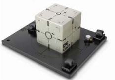
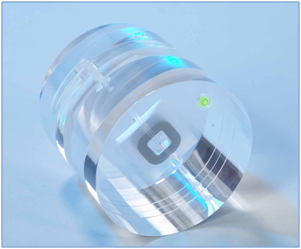
112B Focal Spot Test Tool
Target positioning through imaging guidance is critical to the accurate delivery of radiation treatment. Verifying that all of the imaging, localization and targeting systems are aligned with the true radiation isocenter is crucial. The ISO Cube provides a cost-effective, quick and accurate means of testing radiation isocenters of the imaging guidance systems.
The ISO Cube was designed specifically for daily system checks. The lasers and light field can be turned to the true radiation isocenter using the engraved markings on the exterior of the ISO Cube. The light field and radiation field alignment can be checked using integral radiographic markers. More importantly, the isocenters of both the ODI and the EPID can be checked for true spacial alignment and co-incidence with that of the treatment beam.
The ISO Cube contains a unique center point fiducial and an offset target. The offset target is used to insure the table offset co-ordinates generated by kV/MV imaging are accurate by locating the target, moving the table to the determined amounts and verifying that the offset target has been positioned at the isocenter. The exterior is machined with concentric circle targets to allow the user to objectively assess all set-up errors, including rotations, and to easily align the phantom to the true radiation isocenter.
665 Series
Acrylic Calibration Check Phantoms
These economical calibration check phantoms are solid 15cm cubes, constructed of laminated acrylic. Photon calibration checks can be conveniently and quickly performed at 5cm depth through the top surface. The phantom is simply inverted for photon measurements at 10cm utilizing same chamber cavity. The 665-600 and 665-700 series calibration check phantoms provide additional electron measurement at 1cm and 1.5cm depth, respectively, with an additional chamber cavity on one side. Field markings of 10cm² are provided on all beam entrance surfaces.
Model 458
151 Low-Resolution Test Tool
The Model 458 Calibration Check Phantom is a quality control test tool constructed of Solid Water®, designed to be used for energy checks and calibration of megavoltage machines in a radiotherapy department. The epoxy resin-based Solid Water® material has attenuation characteristics equivalent to water for both electrons and photons.
The Solid Water® Calibration Check Phantom consists of a slab with six cavities and six matching positives made of Solid Water®material which serve to plug the cavities that are not being used. An additional positive is provided which is constructed to fit a cylindrical ionization chamber specified at the time of purchase of your Model 458 Calibration Check Phantom to serve as a chamber adapter.
The six cavities with Solid Water® plugs are located at depths of 1.2cm, 1.5cm, 2.0cm, 2.5cm, 3.2cm and 5.0cm allowing measurements at dmax for photon beams. Each plug can be replaced with the Solid Water® chamber adapter and ionization chamber combination, allowing measurements at a specific depth while the other Solid Water® plugs remain inserted so that the phantom is a consistent slab of Solid Water®. When this is done, the reference point of the ionization chamber will be located under the appropriate center cross hair on the surface of the phantom slab. The 10 x 10cm squares inscribed on the slab are used to set your light field.
All six Solid Water® cavity plugs may be used to make the Solid Water® Calibration Check Phantom a solid piece of material, and the conveniently selected 30 x 30cm size allows this phantom to be used in place of a standard 6cm thick slab within a calibration phantom assembly. To enable the use of a second ion chamber of a different design, only an additional chamber adaptor custom drilled to match the chamber dimensions needs to be purchased.
Model WP-5230
Water-Filled Calibration Phantom
The CNMC Model WP-5230 is an economical, convenient, easily transported acrylic water-filled calibration check phantom. It is compact, measuring only 20 x 20 x 10cm and weighing just under 3 lbs when empty.
The entrance window is engraved with 10cm field and central axis markings. An acrylic transverse-mounted sleeve supports any Farmer-type ion chamber, including the Capintec PR-06C. The ion chamber’s reference point is automatically aligned with the field central axis engraved on the entrance window. The chamber depth is fixed at water-equivalent 5cm. As such, the WP-5230 water phantom provides good characteristics for routine checks of the beam output from 2 to 10MV photons.
A screw plug can be removed for easy filling of the phantom with water; boiled, distilled water is preferred. The plug is designed so the phantom will be fully water-tight at any angle. The phantom is fitted with two expansion vessels to cover a temperature range of 5 – 35ºC, extending the practical range of both temperature and pressure.
Model SWP-1427
Stereotactic Calibration Phantom
The CNMC Model SWP-1427 water-filled stereotactic calibration phantom offers an economical means of achieving the desired depth when calibrating a stereotactic beam with a N23342 plane-parallel ion chamber. The phantom consists of a 13cm diameter acrylic cylinder, 27cm high, bonded to a 14cm x 14cm, 5mm thick water-equivalent plastic base. This configuration offers zero depth correction. The water level may be conveniently adjusted using the spigot mounted in the side of the cylinder.
In use, the N23342 ion chamber is placed into a phantom section that is custom machined to position the chamber sensitive volume at the exact beam central axis, with its window flush with the phantom top surface. The water-filled calibration phantom is then placed on top of the phantom section. Exact depth is achieved by adding water to the cylinder.
In the same manner, the SWP-1427 may be used on top of the STP-156 phantom that accommodates a small field semiconductor detector for achieving variable build-up.
Model LIU-0312
Liu Calibration Check Phantom
The Model LIU-0312 Calibration Check Phantom is a convenient, water-filled acrylic phantom designed to be used for output checks and calibration of megavoltage machines in a radiotherapy department. This economical phantom can be used for both photon and electron beam QA.
The LIU Phantom is an acrylic cube with three custom-made ionization chamber holders for your particular chamber. Two small filler holes are in place to facilitate the filling and emptying of the phantom with water. A crosshair and the outline of a 10cm x 10cm field are inscribed on the four windowed walls.
The three-chamber holder cavities are located at depths of 1cm, 2cm, 5cm, and 10cm, allowing measurements at dmax for photon beams. The chamber holder cavities are strategically located so that no cavity interferes with another cavity during measurements.
The LIU phantom allows for chamber placement to be consistent from week to week during measurements. Quick laser alignment checks can also simultaneously be performed.
Model 670
Head Scatter Mini-phantom
The Model 670 water-equivalent, cylinder mini-phantom improves the dosimetric accuracy and reliability of the linear accelerator beam MU calculation by taking into account stray scatter radiation and electron contamination when determining the total radiation dose given. Head scatter, the stray radiation that bounces around inside the head of a linear accelerator, occurs with each machine. Some of this radiation is scattered forward and hits the patient to become a contributing factor to the total radiation dose.
Application of this phantom is described in ESTRO Booklet March 1997, “Monitor unit calculation for high energy photon beams" for output, volume scatter and scatter primary ratio measurements. By making a measurement with the Miniphantom at a reference depth of 10cm, the physicist can investigate and isolate the influence of scatter radiation on a reference dose measured in a slab phantom.
The Mini-phantom consists of a cylinder, 4cm in diameter, 17.5cm long, made of water-equivalent Plastic Water®. An alignment groove scribed at 10cm serves to indicate the isocenter and coincides with the ion chamber reference point. The standard chamber cavity, parallel to the phantom axis, is for the classic Farmer-type chamber, but it can be made for any commercially-available cylindrical chamber at no additional cost. The Mini-phantom may be used with the CNMC Model AL-CSSP chamber stand equipped with a special vertical adaptor or any suitable chamber stand that allows vertical chamber orientation.
Model AL-CSSP
Identical to the Model AL-CSS chamber stand, but it includes an additional 90° quick-release clamp to vertically position a Farmer-type chamber or any cylindrical chamber with a stem of up to 12.7mm diameter. It retains the ability of the Model AL-CSS to hold ion chambers in a horizontal orientation for in-air measurements.
Plastic Water® is a registered trademark of Computerized Imaging Reference Systems, Inc. (C.I.R.S.)
Anthromorphic
ART Phantoms
for Radiation Treatment Planning
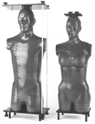
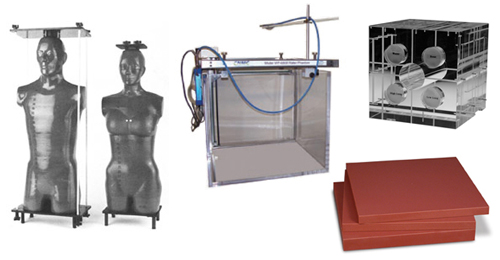
Because of variations of natural human skeletons, unknown calcium loss (approaching osteoporosis in some cases) and contamination by bleaches and other chemical agents in bone preparation, the ART “Superhuman" skeletons are designed and constructed to be both realistic and consistent. Molds for both the cortical bone and the medullary cavities were made using natural skeletons. Bone uniformity derived by this method acilitates positioning within the soft tissues, eliminating the need to make compromises and modifications, as must be done with natural bones, to fit within fixed molds. Complete average-sized male and small-sized female phantoms may be assembled externally for film dosimetry or, internally, for TLD. Plates and tie rods for both assembly methods are included with every phantom.
Soft tissue materials are matched to muscle in specific gravity, mass density and absorption coefficients. Lungs are molded from syntactic foam, with a specific gravity of 0.30.
The ART phantom is transected horizontally in 1" slices and may be ordered undrilled for film dosimetry or drilled for TLD. The drilled phantoms are supplied with pins of appropriate material that may be replaced with optional TLD holders. Holes are available in 5 or 7 mm diameters on 1.5 x 1.5 cm or 3 x 3 cm grids.
Breasts can be ordered in various sizes. They can be drilled in the AP direction for dosimetry or sliced in frontal planes (drilled or undrilled) for film dosimetry. The male chest with breasts attached serves as a large female.
Nylon rods for internal assembly pass through registration holes, held by aluminum assembly plates, and clamped by knobs at the ends. The external assembly plates are larger, allowing tie rods to remain external to the larger phantom contours.
Solid pins are provided with drilled phantoms in appropriate quantity and materials. TLD holders are available in bone, tissue or lung-equivalent material and in 5 or 7mm O.D.
Break-Apart PIXY
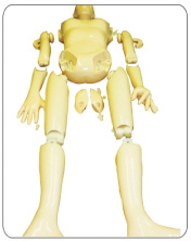
RS-103 or RS-103T
Contains fill ports for the stomach, gall bladder, urinary bladder, right and left kidneys, rectum and sigmoid flexer. Includes permanent storage case.
RS-104 or RS-104T No fill ports for the stomach, gall bladder, urinary bladder, right and left kidneys, rectum and sigmoid flexer. Includes permanent storage case.
RS-105 or RS-105T No organs, no fill ports. Includes permanent storage case.
Stereotactic
RSVP Phantom™
Radiosurgery Verification Phantom
The RSVP Phantom™ was developed to provide stereotactic localization and dose verification for radiosurgery machines. The phantom may be used for a variety of radiosurgery applications, including periodic quality assurance evaluations and acceptance testing. In addition, the phantom may be used to perform re-evaluations after equipment and software upgrades.
The phantom’s design provides full simulation of the localization and irradiation sequences. The anatomically accurate head form is filled with water to simulate the radiation absorption and scatter of human soft tissue. The heavy-duty outer shell is designed to accommodate the anchoring screws.
An internal container called a tumor vessel can be positioned anywhere within the head form by manipulating an external position rod. This vessel may be filled with a radiation-sensitive gel for alignment evaluations or with TL dosimeters for quantitative dose measurements. An Exradin Microchamber may also be used with a special optional chamber holder and ball assembly.
Stereotactic Localization – An agarose gel that changes color after exposure to a minimum radiation dose of 40 Gy is used. A small radio-opaque target is placed inside the gel. Next, the tumor vessel is positioned within the head form and the radiosurgery head frame is attached to the phantom. The standard protocols are followed to locate the x, y and z coordinates of the patient’s tumor through angiography, CT, or MR imaging. The phantom is then mounted in the radiosurgery system and irradiated. The gel is carefully extracted, sliced and analyzed to determine whether the location and shape of the irradiated portion of the gel corresponds to the desired target location.
Dose Measurements – Either TLD or radiochromic film may be used with appropriate holders to quantify the radiation dose. The phantom can also accommodate a specially designed chamber holder for the Exradin Microchamber Model A14.
Construction – The shell of the RSVP Phantom™ is formed from a transparent, 1/4" cellulose acetate butyrate sheet, chosen for its strength and low water absorption. The shell is mounted on a polycarbonate end plate, and the tumor port and cover plate assembly are attached with nylon screws to the end plate. The cover plate is removable for internal access. The tumor vessel is attached to the external position rod. After the desired position is reached, the vessel is locked into place by hand-tightening a lock nut on the rotation ball and a lock bolt on the position rod. The RSVP Phantom™ includes three tumor vessels and a wooden storage case.
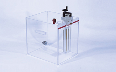
Model STP-145M
Radiosurgery Verification Phantom
The CNMC Model STP-145M combines the STP-145 phantom assembly with a crank-operated, linear-drive mechanism, allowing precision off-axis positioning of the detector.
The phantom assembly slides smoothly on a Teflon® platform, driven by a stainless steel lead screw and stabilized by two stainless steel rods. Each full turn of the hand crank quickly and accurately moves the phantom assembly 1mm. A 5-digit mechanical counter with pushbutton reset capability indicates position to the nearest 0.1 mm. Total detector travel is 16cm or less.
The STP-145M has adjustable feet for leveling the unit. Optional brackets to mount the assembly to a Zemed Danek Linac Scalpel stereotactic radiosurgery system are available.
The STP-145M may be conveniently retrofitted with the optional Model RMD-100-5 and RMD-200-5 remote motor drives that allow detector positioning from a remote location. The detector position is indicated by a 5-digit LCD display with pushbutton zeroing. The convenience of the Model RMD-100-5 and RMD-200-5 provide significant savings to your data acquisition time.
Plastic Slab Phantoms
Acrylic & Polystyrene
Phantom Materials
A typical acrylic or polystyrene phantom is a 25cm cube consisting of one of each fractional thickness, eight 1" thick sections and one 1" thick section which has an ion chamber cavity drilled at 1cm from the nearest surface. This allows the calibration depth to be adjusted in 0.8mm (1/32") increments over 1cm for photon beams. A section for a plane-parallel chamber is machined to position the chamber flush with one surface. In this manner, depths can be achieved for calibrating electron beams in 0.8mm (1/32") steps.
Economy is achieved by using standard thickness material in English System dimensions. Although the thickness tolerance can vary ±5%, the thickness of each section is individually measured and marked with permanent ink in metric units.
Separate sections are available for adding to an existing phantom or to assemble a phantom with custom dimensions.
Polystyrene
Polystyrene plastic (PS), also known as polystyrol, has a density of 1.05g/cm³, which makes it lighter than acrylic. Historically supplied in clear sheets, polystyrene is currently only supplied in milky white sheets. It is available in 25 x 25cm sections of varying thickness.
Acrylic
Acrylic phantom material is a clear plastic with the chemical formula (C-5/H-8/O-2)n, polymethylmethacrylate (PMMA). It is also known under the trade names Lucite, Plexiglas and Perspex. Acrylic has a density of 1.185g/cm³. It is available in 25 x 25cm sections of varying thickness.
Plastic Water®

Phantom Materials
Plastic Water® is resilient and will not break easily under impact, it is easily machined and is the only calibration material available in 1mm thickness. The advanced casting methods assure void-free product, available in two formulations.
Plastic Water® – the Original
For general calibration work, the original Plastic Water® is the only calibration material which agrees with true water within +0.05%/-0.4% above 4MeV (+/-0.5% from 150keV to 100MeV).
Plastic Water® DT
This improved Dignostic/Therapy formulation is optimized to cover CT energies for radiation planning and dose verification in IMRT. It mimics true water within +/-0.22% from 80keV to 4MeV (+/-0.5% from 50keV to 25MeV).
Size Availability
Although 30 x 30cm slabs are most widely used and therefore considered to be a standard size, Plastic Water® is also available in 20 x 20cm, 25 x 25cm and 40 x 40cm sizes.
Chamber Cavities
For dosimetry measurements, custom cavities are available to accommodate any ion chamber on the market today in slabs of any size and thickness of 2cm and over. Matching positives (plugs) are also available for most ion chamber types. The table shows most of the popular ion chambers. Simply add the appropriate suffix to the model number from the table at left.
Plastic Water® is a registered trademark of Computerized Imaging Reference Systems, Inc. (C.I.R.S.)
Virtual Water™
Phantom Materials
Virtual Water™ makes routine checks of your linear accelerator easier. Designed for photon and electron beam calibrations, it eliminates the inconvenience of transporting, setting up and filling water tanks. Virtual Water™ is free of air and other imperfections and is not affected by humidity or temperature changes. Skillfully molded and accurately machined in standard dimensions, Virtual Water™ can help you achieve calibrations within 0.5% of the true dose.
Virtual Water™ scatters and attenuates diagnostic and radiotherapy range X-rays the same way as water without the charge storage problems. It can be used for both photon and electron beam calibrations, including relative ionization, depth dose measurements and absolute calibrations without the need for correction and scaling factors. Ionization readings obtained in Virtual Water™ are practically the same as those in liquid water for the same depth and exposure duration.
Each batch of Virtual Water™ is tested at an independent calibration lab and verified to be within 0.5% of water at photon energies. Although 30 x 30cm slabs are most widely used and therefore considered to be a standard size, Virtual Water™ is also available in various thicknesses in 20 x 20cm and 40 x 40cm sizes.
Virtual Water™ is a trademark of Med-Cal, Inc.
Solid Water®
Phantom Materials
Solid Water® makes radiation beam calibration easier than ever. Designed for photon and electron beam calibrations, it eliminates the inconvenience of transporting, setting up and filling water tanks. Carefully molded and accurately machined into standard dimensions, Solid Water® can help you achieve calibrations within 1% of the true dose.
Solid Water® scatters and attenuates diagnostic and radiotherapy range X-rays the same way as water without the charge storage problems. It can be used for both photon and electron beam calibrations, including relative ionization, depth dose measurements and absolute calibrations without the need for correction and scaling factors. Ionization readings obtained in Solid Water® are virtually the same as those in liquid water for the same depth and exposure duration.
Although 30 x 30cm slabs are most widely used and therefore considered to be a standard size, Solid Water® is also available in various thicknesses in 20 x 20cm and 40 x 40cm sizes.
CTG (Certified Therapy Grade) Solid Water® is a premium product, manufactured to the most exacting quality standards. It is supplied with a Certificate of Conformance which includes the calculated elemental composition, calculated mass and volume electron densities, electron and photon transmission characteristics and measured physical dimensions. A radiograph of each slab demonstrates the product is free from voids, contamination and other artifacts. It is available in all sizes and thicknesses. Simply add the suffix “-CTG" to the indicated model numbers.
SolidWater® is a registered trademark of Gammex RMI
Water Phantoms
Water Phantoms
with Depth Positioning Assembly
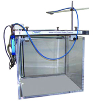
The CNMC Models WP-3040 and WP-3840 are water phantoms with convenient manual ion chamber depth positioning. The water tanks are constructed of 3/8" acrylic and provided with side-mounted handles, a drain and a ball valve.
The outstanding feature is the depth positioning assembly that is common to both models and is unique in construction and operation. The ion chamber holder Teflon® block slides on two stainless steel rods and is driven by a stainless steel lead screw. One turn of the hand crank quickly and accurately moves the chamber holder block 1mm. A mechanical counter with push button reset capability indicates depth to the nearest 0.1mm.
The chamber holder block has a 7mm hole with 3 set screws, designed to hold various chamber holders. The chamber holder that is supplied with the phantom can accommodate cylindrical ion chambers of various diameters, from 3mm to 16mm. Plane-parallel ion chamber holders are available as an option to accommodate Markus® or NACP chambers and Roos® chambers.
The horizontal off-center positioning is achieved by slicing the depth positioning assembly along the edge of the water tank to the desired position as indicated on the horizontal scale mounted on the end of the tank. The larger phantom features an additional scale on the side of the tank.
The depth positioning assembly of both models may be easily retro-fitted with an optional Remote Motor Drive that allows chamber depth positioning from a remote location. The depth is indicated by a digital LCD display with push button zeroing, adding convenience and significant time saving to your monthly routine.
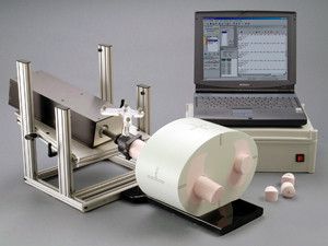
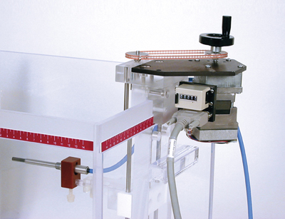
Model WP-6868
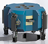
3D Radiation Scanning System
The 3D Radiation Scanning System is fast, accurate, simple, and easy to set up. A RS-485 network cable is the only link with the outside computer. The scanners are constructed with accuracy, simplicity and portability as priorities.
Features
- Interfaces with all major treatment planning systems
- Accuracy of positioning is 0.1 mm or less
- Accuracy of measurement is 0.5% or less
- Easy in-room setup with iPad (included)
- Network cable is the only link to the outside computer
- iPad pendant
- Made of clear anodized machined aluminum
System Requirements
Recommended system requirements for optimally running the WP-6868:
- 2 GB of RAM
- 100 MB Hard drive space
- SVGA with 32-bit true color
- Windows based operating system (Vista or later)
- Minimum screen resolution: 1024 x 768
Virtual Water™
Remote Motor Drive
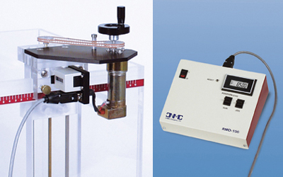
With the CNMC Model RMD-100-5 Remote Motor Drive you can convert the manual mechanism of the CNMC WP-3040, WP-3840 and STP-145M detector positioning assembly to allow detector positioning from outside of the treatment room, adding convenience and significant savings to your data acquisition time.
Installation is easy, since it makes use of existing threaded holes for mounting the motor drive assembly to the detector positioning assembly. Although the installation includes adding two drive sprockets, the hand crank is reinstalled on the motor shaft, thereby retaining the original manual positioning and mechanical counter readout functions.
The control/display module is connected to the drive motor by a 15-meter cable for complete control of detector position from the accelerator console. Its six-digit liquid crystal display indicates the depth position in units of centimeters to 0.001cm. The RUN switch operates the motor drive in a continuous mode, while the JOG switch provides intermittent operation. Both switches control the motor drive in up/down directions, indicated on display as + and – respectively.
Model RMD-100-1 may be specified for adaptation to the existing Med-Tec MT-100 and MT-150 water phantoms. Except for the mounting hardware, all features and technical specifications are indentical to the RMD-100-5.
Model RMD-100-3 may be specified for adaptation to the existing CNMC WP-300 and WP-380 water phantoms. Except for the mounting hardware, all features and technical specifications are indentical to the RMD-100-5.
Model RMD-200-5-PC
Phantom Materials
With the Model RMD-200-5-PC, it is possible to convert the manual, crank-operated depth-positioning mechanism of the CNMC WP-3040 and WP-3840 water phantoms to allow ion chamber positioning from outside the treatment room, using a PC (personal computer) as the control panel, thus adding convenience and significant savings to your monthly routine.
Installation of the motor drive assembly to the depth position mechanism is easy, since it makes use of existing threaded holes. Although the installation includes adding two drive sprockets, the hand crank is reinstalled on the motor shaft, retaining the original manual positioning and mechanical counter readout functions.
The PC software serves as the control/display unit. It is connected to the drive motor by a 15-meter cable via an interface unit for complete control of chamber depth from the accelerator console. The programmability of the PC allows the user to present the depth increments or the entire measurement routine. This display indicates the depth position, with 0.01cm accuracy assured by optical encoder feedback.
The PC Drive can be supplied with motor mounting hardware for adaptation to water phantoms of older design. All features and technical specifications remain the same.
Model RMD-200-1-PC may be specified for adaptation to the existing MEDTEC MT-100 and MT-150 water phantoms.
Model RMD-200-3-PC may be specified for adaptation to the existing CNMC WP-300 and WP-380 water phantoms.
Model RMD-200-5-PC
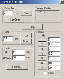
Software for Remote Motor Drive
With the Model RMD-200-PC software, it is now possible to control your RMD-200-X remote motor drive from your PC or MAC from outside of the treatment room, thus adding convenience and significant time saving to your monthly routine. Installation of the software is easy. Just copy the .exe file and go. The hand crank is reinstalled on the motor shaft, retaining the original manual reference point positioning and mechanical counter readout functions.
The PC or MAC computer along with the CNMC motion control software serves as the control/display unit. It is connected to the drive motor by a 15 meter cable via an interface unit for complete control of chamber depth from the accelerator console. The programmability of the softwre allows the user to preset the depth increments or the entire measurement routine. The computer display indicates the depth position, with 0.01cm accuracy assured by optical encoder feedback.
Model RMD-200-1-PC may be specified for adaptation to the existing MEDTEC MT-100 and MT-150 water phantoms.
Model RMD-200-3-PC may be specified for adaptation to the existing CNMC WP-300 and WP-380 water phantoms.
Model RMD-200-5-PC may be specified for adaptation to the existing CNMC WP-3040 and WP-3840 water phantoms.
Universal Lift Carriage
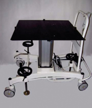
Our Universal Lift Carriage is designed to accurately position large water phantoms when critical measurements need to be made. This high-precision electromechanical lift carriage has a full 3 inch floor clearance, 4 large diameter wheels, an in independent manually operated braking system to simultaneously lock all 4 wheels and almost 16 inches of vertical movement.
Waterproofing Devices
for Farmer-type Chambers
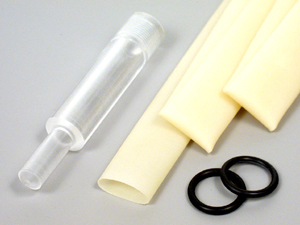
Various waterproofing kits have been developed by CNMC to effectively waterproof a Farmer-type 0.6cc dosimetry ionization chamber for use in water phantoms. Their unique design has a distinct advantage over rubber or latex sheaths by eliminating the possibility of talcum powder contamination.
The heart of the design is an acrylic cap that has been machined down to 1mm wall thickness (compatible with TG-51 protocol) at the chamber thimble area. This housing assembly is designed to accept a Farmer-type ion chamber. A flexible tube fits snugly over the open end of the cap, and the addition of two rubber O-rings insures a watertight seal.
Since various manufacturers’ chambers may exhibit minor physical differences, CNMC offers several versions to accommodate most chambers.
As with latex and rubber sheaths, the flexible tubing may harden over time and should be inspected before use and replaced periodically. However, this tubing is relatively inexpensive and is readily obtained from CNMC or through the hospital’s surgical supplier.
WPK-75 for NE Chambers
This set consists of an acrylic cap with M11x.75 thread which fits NE Farmer-type chambers, six 5/8" diameter Penrose drainage tubes and two 5/8" diameter O-rings.
WPK-75C for Capintec PR-06C
This set consists of a larger 3/4" acrylic cap with M11x.75 thread, six 3/4" diameter Penrose drainage tubes and two 3/4" diameter o-rings to accommodate the cable connector of the PR-06C. It also fits NE Farmer-type chambers.
WPK-100 for PTW Farmer® Chambers
This set consists of an acrylic cap with M11x1 thread which fits all PTW Farmer-type chambers, six 5/8" diameter Penrose drainage tubes and two 5/8" diameter O-rings.
WPK-691 Universal Waterproofing Kit
This set contains a uniquely designed acrylic waterproofing cap without the threads. An internal O-ring with air bypass holds all Farmer-type ion chambers securely, including the Capintec PR-06G and PR-06C. The set includes six 3/4" diameter Penrose drainage tubes that can accommodate the cable connector of the PR-06C without the need for machining.
2513A Waterproofing Sheath
These latex sheaths are molded to conform to all outside dimensions of PTW, NE and Capintec PR-06G Farmer-type ion chambers. To keep the inside walls from sticking while in storage, the sheaths are supplied dusted with talcum powder. The sheaths are more difficult to install, remove and store, but they can be used successfully to waterproof Farmer-type chambers providing that extreme caution is taken to prevent talcum powder contamination of the chamber.
PTW Farmer® is a registered trademark of PTW Freiburg and PTW New York.